Embolization of renal cell carcinoma metastasis
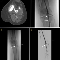
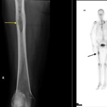
Renal cell carcinomas tend to be highly vascular tumours, and any RCC metastases that develop tend to be similar. This patient presented several years after nephrectomy for RCC with a solitary painful bone metastasis (image (a), arrows). Resection, cementing and plate fixation were planned by the orthopaedic team however it was considered likely that there would be massive blood loss at the time of surgery due to the hypervascular nature of the metastasis. Pre-operative embolisation was requested. At angiography, a large branch of the popliteal artery (image (b), labelled ‘P’) was identified feeding the tumour, which looks very vascular (arrows). This branch was embolized with coils (image (c), arrow), following which no contrast was identified filling the tumour. The patient was brought straight to surgery and resection of the lesion was uneventful. Post-operative appearance is shown in image (d), with high density cement filling the defect left by curettage of the metastasis.


