Giant Cell Arteritis – CT
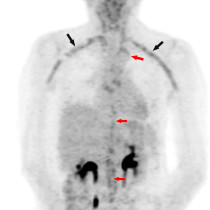
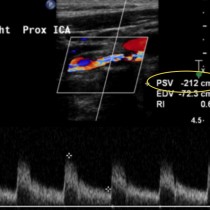
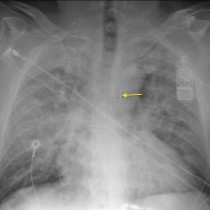
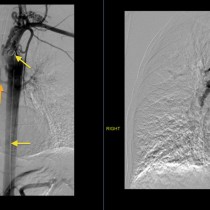
Giant cell arteritis. This 73 year old woman presented with headaches. ESR and CRP were elevated and GCA was suspected. This CT angiogram shows circumferential thickening of the walls of the left subclavian and common carotid arteries (arrows), as well as the descending thoracic aorta (arrowheads), typical of arteritis. The diagnosis was confirmed on temporal artery biopsy.


