Global hypoxic ischaemic injury
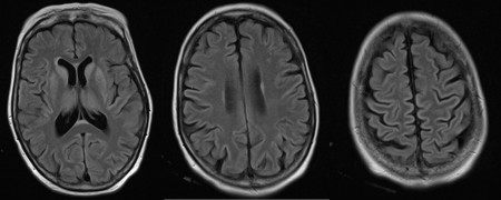
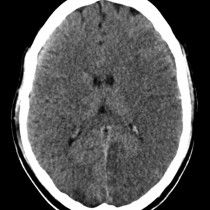
Global hypoxic ischaemic injury. This patient suffered an out-of-hospital cardiac arrest and was subsequently resuscitated, however failed to wake up. These three images are from the FLAIR sequence of the patient’s MRI and are grossly abnormal, however the abnormality is subtle because it is so diffuse and symmetric. Note how prominent the cortical grey matter and the basal ganglia are – they are abnormally hyperintense because of cytotoxic oedema. Grey matter is much more sensitive to ischaemia, therefore in global hypoxic ischaemia injury imaging will show abnormal appearance of the cortex, basal ganglia, thalami and cerebellum. This may be visible on CT (as abnormal low attenuation grey matter) however MRI is far more sensitive, particularly FLAIR and diffusion weighted imaging.

