Graves disease
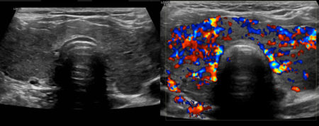
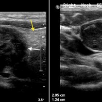
This 30-year-old woman presented with classic symptoms and biochemical evidence of thyrotoxicosis, and was referred for a thyroid ultrasound.
The grey-scale image on the left shows a diffusely abnormal-appearing thyroid gland; it is enlarged, heterogeneous and hypoechoic. The Doppler image on the right shows diffusely increased vascularity throughout the thyroid. This appearance is called a ‘thyroid inferno’. These are the typical sonographic features of Graves disease.
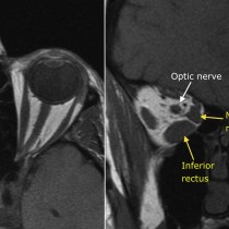
Graves disease is frequently associated with some extra-thyroid manifestations, the most common of which is thyroid-associated orbitopathy (see an example of this here), whereby some or all of the extra-ocular muscles become enlarged, resulting in proptosis. Other manifestations are thyroid dermopathy (pretibial myxoedema), and thyroid acropachy (rare, causing finger clubbing and periosteal reaction along the tubular bones of the hands and feet).
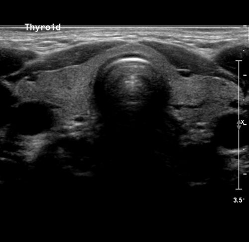
The normal thyroid is uniformly hyperechoic on ultrasound – an example of this is provided below for comparison.
