Occluded middle cerebral artery on CT angiography
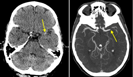
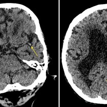
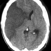
This 69 year old man presented to ED with acute right hemiparesis. He was immediately referred for a CT brain and, as he was within the window for potential thrombolysis, also for CT angiography. The CT brain showed no parenchymal abnormality (which is typically the case in the early stages of an acute stroke) and no intracranial haemorrhage, however there is a good example of the dense middle cerebral artery (MCA) sign (left image, arrow). This sign suggests MCA thrombosis, but is not specific.
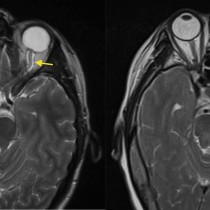
With the advent of newer therapeutic options for stroke such as intra-arterial thrombolysis and thrombectomy, CT angiography has now become a routine diagnostic tool in the work-up of these patients. The purpose of CT angiography in the setting of an acute stroke is to assess the potency of the carotid, vertebral and intracranial arteries. It will show the site of any occlusion, delineate the extent of atherosclerotic disease, and demonstrate dissection of vessels if present. In this case, the CT angio image on the right confirms that there is complete occlusion of the left MCA (arrow).

