Osteoid osteoma
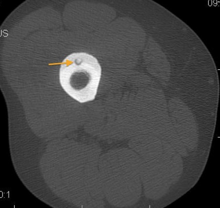
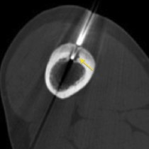
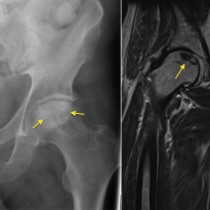
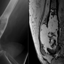
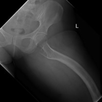
Osteoid osteoma. This CT image from the femur of a young man shows a small lytic lesion with a central density located in the anterior cortex of the femur (arrow), associated with a thick rind of new bone on the surface of the cortex (slightly less dense than the native cortex). Appearances are typical for an osteoid osteoma. These are very painful lesions, but the pain is characteristically relieved by aspirin. Nowadays, they are treated by radiofrequency ablation under CT guidance.

