Portacath insertion
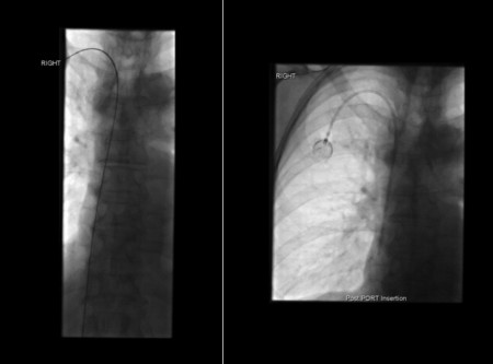
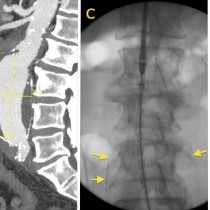
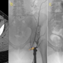
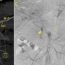
Portacath insertion. The most common indications for portacaths include oncology and, in SVUH, cystic fibrosis patients. A guide wire is first inserted, through one of the internal jugular veins and then along the SVC to the right atrium (left-hand image). The catheter is then advanced over the guidewire, and attached to the port which is positioned subcutaneously in the chest wall (right image). If you look very carefully, you will notice that you can read the letters ‘CT’ on the port – this is to indicate that the device is capable of being used for high-pressure pump-driven injections of contrast, as typically used in CT studies. Portacaths have been revolutionary for the patients who need them, as they can be used for drug administration, blood tests, and contrast injections, saving them dozens of venipunctures over the course of their treatment.

