Scaphoid fracture
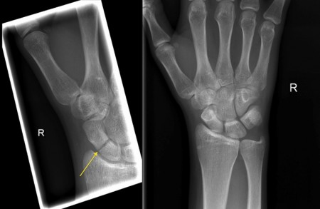
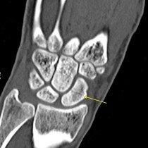
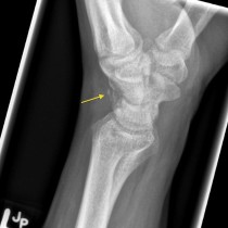
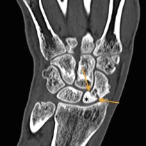
Scaphoid fracture. The image on the left clearly shows a transverse fracture of the scaphoid waist. This radiograph is a dedicated ‘scaphoid view’, a technique which elongates the scaphoid and makes it much easier to see fractures. To illustrate this, the image on the right is from the same patient, taken at the same time, and is a standard AP view of the wrist – the fracture is not visible. Because these fractures are notoriously difficult to see on initial radiographs, when one is suspected but not seen the patient is brought back for repeat views a week to ten days later, at which time some healing at the fracture site will usually make it more conspicuous.

