Scapular fracture
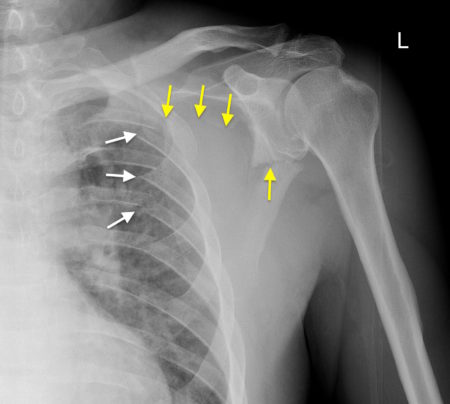
This young man was knocked from his bicycle and was taken to the ED complaining of severe pain around his left shoulder.
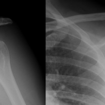
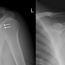
This frontal radiograph of his shoulder shows a displaced fracture through the body of the scapula (yellow arrows).
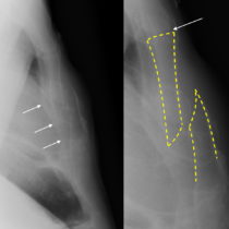
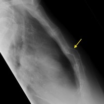
There are also fractures of the posterior aspects of several of the upper left ribs (white arrows).
Scapular fractures are relatively uncommon injuries; the scapula is well-protected from traumatic forces by the chest wall anteriorly, the large muscles that surround it and the fact that it quite a mobile structure. As a result, it generally takes high-energy trauma to fracture it, such as car or motorcycle accidents, high-speed cycling accidents and falling from a height. They have also been reported to occur in patients following a seizure.
Because they make up as few as 3% of shoulder fractures, we see very few of them and they are quite easy to overlook when we are interpreting shoulder radiographs in a trauma setting.
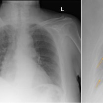
While painful, scapular fractures are not particular severe injuries on their own as the majority are minimally displaced and heal without surgical intervention. However, the importance of identifying a scapular fracture on imaging relates to the fact that, because of the high-energy trauma required to cause them, they are very commonly associated with other injuries which may be life-threatening. These include rib fractures, as in this case, as well as clavicle fractures, pneumothorax and pulmonary contusions, vertebral fractures, and intracranial haemorrhage.
The majority of scapular fractures involve the body or neck. Less commonly, the spine, acromion or coracoid process of the scapula can fracture. A small proportion of scapular fractures involve the glenoid and are therefore intra-articular – these are more likely to require internal fixation.
Reference: Ropp AM, Davis DL. Scapular fractures: what radiologists need to know. AJR 2015;2015:491-501. doi: 10.2214/AJR.15.14446

